Tag Archives: Chiropractic

Examination of Lymph Nodes.

Brief Examination of the Lymph Nodes


The lymphatic system is a series of vessels throughout the body that drain fluid from tissues.  Bacteria and other microbes are picked up in the lymphatic fluid and trapped inside the lymph nodes, where they can be attacked and destroyed by white blood cells.  Common problems of the lymphatic system include Glandular Fever, Hodgkin’s disease, Oedema and tonsillitis. 


Sequence:  Inspect the patient for a visible lymphadenopathy  (any disease of the lymph nodes). Afterwards, palpate the patient one side at a time, whilst comparing with the nodules on the contralateral side. 


Assessment: 

  • Site.
  • Size. 
  • Consistency.
  • Tenderness.

One major side is on the left supraclavicular fossa (the seat of the Devil) – Virchow’s Node – Linked to abdomen cancer.  Known as Troisier’s sign. Suspect this if the node is hard and enlarged above the average (0.5cm) and the max being 2.0cm. Fixation of lymph nodes also points towards malignancy.  nejmicm1204740_f1 Consistency – Normal nodes feel soft. They can also feel tender (acute viral or bacterial infection).   It is important to know that sometimes it is natural to have naturally large lymph nodes in some individuals. Proceed with caution anyway. position_oflymphnodesLump-under-Armpit LymphNodes 600

Thank you for reading! I hope you found this helpful! If you have anything to add, please let me know!

Like, Share, Comment, Follow.

Cranial Nerve Examination

Full Cranial Nerve Examination For OS4.

Cranial Nerves


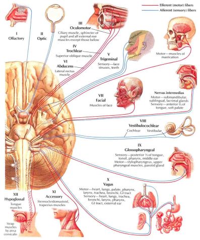
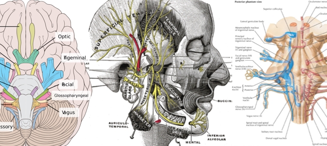
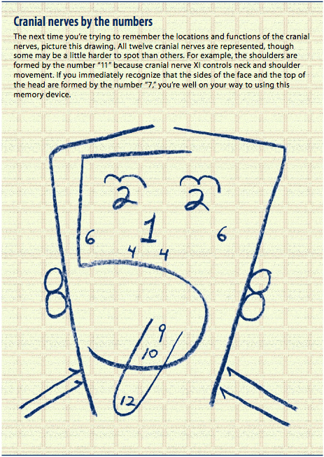
The 12 Cranial Nerves are:  
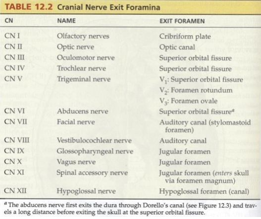
  • I Olfactory.
  • II Optic.
  • III Oculomotor. 
  • IV Trochlear.
  • V Trigeminal. 
  • VI Abducent. 
  • VII Facial. 
  • VIII Vestibulocochlear. 
  • IX Glossopharyngeal. 
  • X Vagus. 
  • XI Accessory. 
  • XII Hypoglossal.

tumblr_lsau0bTUR91r0l4eno1_500


  • The Cranial Nerves’ Song!! – https://www.youtube.com/watch?v=IBuPzn_8UTc

Cranial Nerve Exit Foramina

Bear in mind that this is a rude way to remember the 12 cranial nerves, but please don’t be offended, its just for memory purposes. There are plenty of ‘clean’ options too on the internet. I use: 

Oh, Oh, Oh, To, Touch, And, Feel, Virgin, Girl’s, Vaginas, Ahh!, Heaven. 


Classification of Cranial Nerve Functions: 

  • Sensory: Contain only afferent fibres: 
  • I Olfactory. 
  • II Optic. 
  • VIII Vestibulocochlear. 
  • Motor: Contains only efferent fibres: 
  • III Oculomotor. 
  • IV Trochlear. 
  • VI Abducent. 
  • XI Accessory. 
  • XII Hypoglossal. 
  • Mixed nerves: Contains both efferent and afferent fibres: 
  • V Trigeminal. 
  • VII Facial. 
  • XI Glossopharyngeal.
  • X Vagus. 

Mnemonic: Again, apologies for being rude, but there are ‘clean’ alternatives on the internet. 

S= Sensory, M= Motor, B = Both. 

  • Skinny Sluts Make,
  • Money But My,
  • Brother Says Big, 
  • Bitches Make More. 
  • I Olfactory, II Optic, III Oculomotor, 
  • IV Trochlear, V Trigeminal, VI Abducent, 
  • VII Facial, VIII Vestibulocochlear, IX Glossopharyngeal, 
  • X Vagus, VI Accessory, XII Hypoglossal.

CN 1 – Olfactory Nerve: 

  • Olfactory bulb origin. 
  • Cribiform plate. 
  • Sensory. 
  • Vulnerable to: 
    • + Fractures. 
    • + Tumours. 
    • + Parkinson’s Disease. Progressive nervous system degeneration of the basal ganglia and a deficiency of the neurotransmitter dopamine. Marked by tremor, muscular rigidity. 
    • + Huntington’s Disease. A hereditary disease. Degeneration of brain cells and causing chorea (Kaw-rea) and progressive dementia (memory disorders, personality changes, and impaired reasoning). 
  • Disruption leads to loss of smell, full or partial. 
  • Examination: 
  • Patient closes one nostril and both eyes. 
  • Present coffee to identify.  

CN 2 – Optic Nerve: 

  • Sensory. 
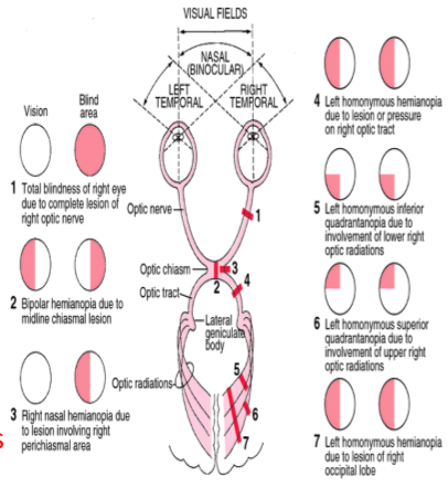
  • Visual pathway from retina, chiasma, optic tracts leading to the visual cortex. 
  • Disruption –> visual disturbances, visual agnosia (can see but can’t interpret the information), disturbances of perception and hallucinations. 
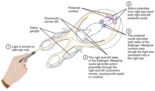
  • How the pupils respond to light: 
  • Direct and consensual response: 
  • The direct response:
    • + Meaning constriction of the illuminated pupil. 
    • + The direct response is impaired in lesions of the ipsilateral optic nerve, the pretectal area (midbrain structure), the ipsilateral parasympathetics traveling in CN III, or the pupillary constrictor muscle of the iris.
  • The consensual response:
    • + Meaning constriction of the opposite pupil. 
    • + The consensual response is impaired in lesions of the contralateral optic nerve, the pretectal area, the ipsilateral parasympathetics traveling in CN III, or the pupillary constrictor muscle.
  • How it works: 
  • Information enters via CN2 – Optic & activates CN3 – Oculomotor.
  • This is a consensual (both eyes are involved) parasympathetic reflex.
  •  If only the eye being tested constricts, then there is damage to the crossing fibres (damage in the midbrain). 

1509_Pupillary_Reflex_Pathways

  • Examination: 
  • Vision chart. 
  • Colour blind test. 
  • Visual reflexes:
    • + Patient covers one eye, shine pen torch into one eye for approx 1 second. – Pupil constriction. 
  • Accommodation: 
    • + Testing for the pupillary response. Normally the pupils constrict  whilst fixating on an object being moved from far to near the eyes. 
    • + Tell the patient to focus on the furthest point of the room, and to focus when adding the pen into their vision. 
  • Accommodation is impaired in lesions of the ipsilateral optic nerve, the ipsilateral parasympathetics traveling in CN III, or the pupillary constrictor muscle, or in bilateral lesions of the pathways from the optic tracts to the visual cortex. 
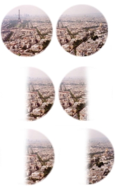
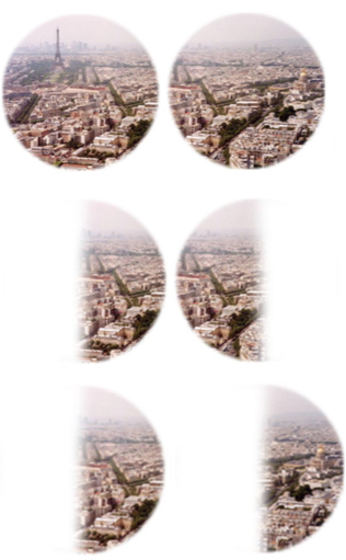
  • Visual field test: 
    • + Ask patient to keep their eyes looking towards your nose. 
    • + In all four quadrants, wiggle your fingers and ask the patient to identify if they see them.
  • Common field defects include: 
  • 1 = Bitemporal Heminanopia: Lesion at the optic chiasm (Pituitary tumour). 
  • 2= Homonymous Heminanopia: Lesion posterior to the optic chiasm (Posterior cerebral artery territory infarction). 
  • 3= Blindness in one eye: Lesion in the eye, retina or the optic nerve. 

Visual Disturbances

Optic Nerve Chiasm


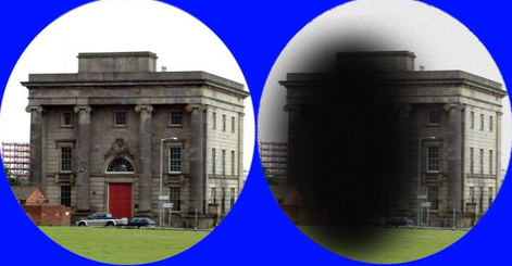
  • Expansion of the blind spot – central scotoma. 
  • Central scotoma is an area of depressed vision that corresponds with the point of fixation and interferes with central vision. It suggests a lesion between the optic nerve head and the chiasm.
    • + Multiple sclerosis – which may cause unilateral or asymmetrical bilateral scotoma
    • + Nutritional causes – which may be due to, e.g. alcohol or tobacco amblyopia, B12 deficiency
    • + Vascular lesions – which may cause unilateral scotoma
    • + Gliomas of the optic nerve – this may cause unilateral scotoma
    • + Simple glaucoma – which may initially cause an off-centre scotoma

Central Scotoma


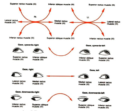
CN III – Oculomotor, CN IV – Trochlear &    CN VI – Abducent: 

  • All involved in movements of the eye. 
  • + Oculomotor: Motor, Oculomotor nucleus, Edinger-Westphal nucleus (both in the midbrain) divides into superior and inferior, through the superior orbital fissure (SOF) and into the orbital cavity.  All other extraocular muscles, also carries parasympathetic (constrictor) fibres to pupil, and fibres to levator palpebrae superioris. 
  •  + Trochlear: Motor, Dorsal Midbrain, SOF, orbital cavity. Supplies superior oblique muscle
  • + Abducent: Motor, Lower Pons, SOF, orbital cavity. Supplies lateral rectus muscle
  • For the full anatomy, see the link for a short powerpoint: http://www.slideshare.net/farhan_aq91/cranial-nerve-iii-iv-and-vi 
  • Examination: 
  • The ‘H’ Test: 
    • + Ask the patient to keep their head in a straight position, whilst using a (preferably red tipped) pen and draw an H shape with their eyes following. 
    • + Ask the patient to report any Diplopia (double vision), if so, at what point. Also keep an eye out for nystagmus (involuntary eye shaking).  
  • http://www.nhs.uk/Conditions/Double-vision/Pages/Causes.aspx
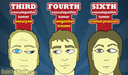
Untitled

Eye Movements: 

  • Look at eyes in a relaxed state. 
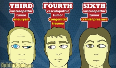
  • 3rd nerve palsy: 
    • + Eye often down and out.
  • 6th nerve palsy: 
    • + Osten eyes convergent (unchallenged medial rectus). 
  • Look at the pupils. – Dilation – bilateral or unilateral? 
  • Look for drooping of the eye lid = Ptosis. Cranial Nerve Disorders

CN V – Trigeminal Nerve:

  • Both sensory and motor.
  • Pons, Medulla and Midbrain origin.
  • Exits: 
    • + V1 = SOF.
    • + V2 = Rotundum. 
    • + V3= Ovale. 
  • Sensory = face.
    • + 3 branches = ophthalmic, maxillary and mandibular. 
  • Motor = mastication = masseter and temporalis. 
  • Examination: 
  • Sensory = cotton wool.
  • + Jaw line.
  • + Cheeks. 
  • + Forehead. 
  • The corneal reflex could also be examined as the sensory supply to the cornea is from this nerve.
  • Do this by lightly touching the cornea with the cotton wool. This should cause the patient to shut their eyelids.
  • Motor = Muscle testing. 
  • Ask the patient to clench their teeth, whilst feeling the temporals and masseter muscles. 
  • Ask the patient to open their mouth against practitioner resistance. 
  • Finally, perform the jaw jerk on the patient by placing your left index finger on their chin and striking it with a tendon hammer. This should cause slight protrusion of the jaw. 

CN 6 – Abducent Nerve: 

  • Tested with CN III & CN IV. 
  • Dysfunction of ipsilateral rectus muscle leads to lateral gaze palsy. 

CN 7 – Facial Nerve: 

  • Motor and Sensory and Para Sympathetic. 
  • Pons. Exits Internal Auditory Meatus & Stylomastoid foramen. 
  • Motor: 
    • + Muscles of facial expression. 
  • Sensory: 
    • + Anterior 2/3rd’s of tongue. 
  • Para Symp: 
    • + Supply to lacrimal, submandibular and sub lingual glands. 
  • Dysfunction:
    • + Bell’s palsy. 
    • +/- Ageusia – Inability to taste.
    • +/- Hypogeusia – Reduced ability to taste. 
  • Examination: 
  • Facial expressions. 
    • + Frown. 
    • + Raise eye brows. 
    • + Smile – teeth showing. 
    • + Blow cheeks out. 
    • + Keeps eyes closed against resistance. 

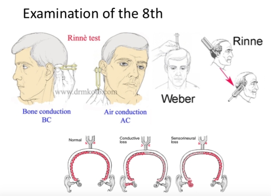
CN 8 – Vestibulocochlear: 

  • Sensory. 
  • Pons and Medula Oblongata. Exits internal acoustic meatus of the temporal bone. 
  • Hearing and equilibrium 
  • Provides innervation to the hearing apparatus of the ear and can be used to differentiate conductive and sensori-neural hearing loss using the Rinne and Weber tests.
  • Conducts two special senses: 
  • Hearing (audition) and balance (vestibular). The receptor cells for these special senses are located in the membranous labyrinth which is embedded in the petrous part of the temporal bone. 
  • Positional vertigo is the most common problem, although tinnitus, hearing loss, and deafness may also occur following damage. Positional vertigo occurs, usually for about 30 seconds, with sudden changes in position, usually from lying to sitting or from sitting to standing. 
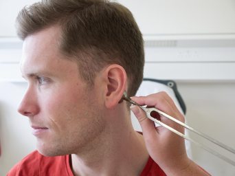
  • Examination: 
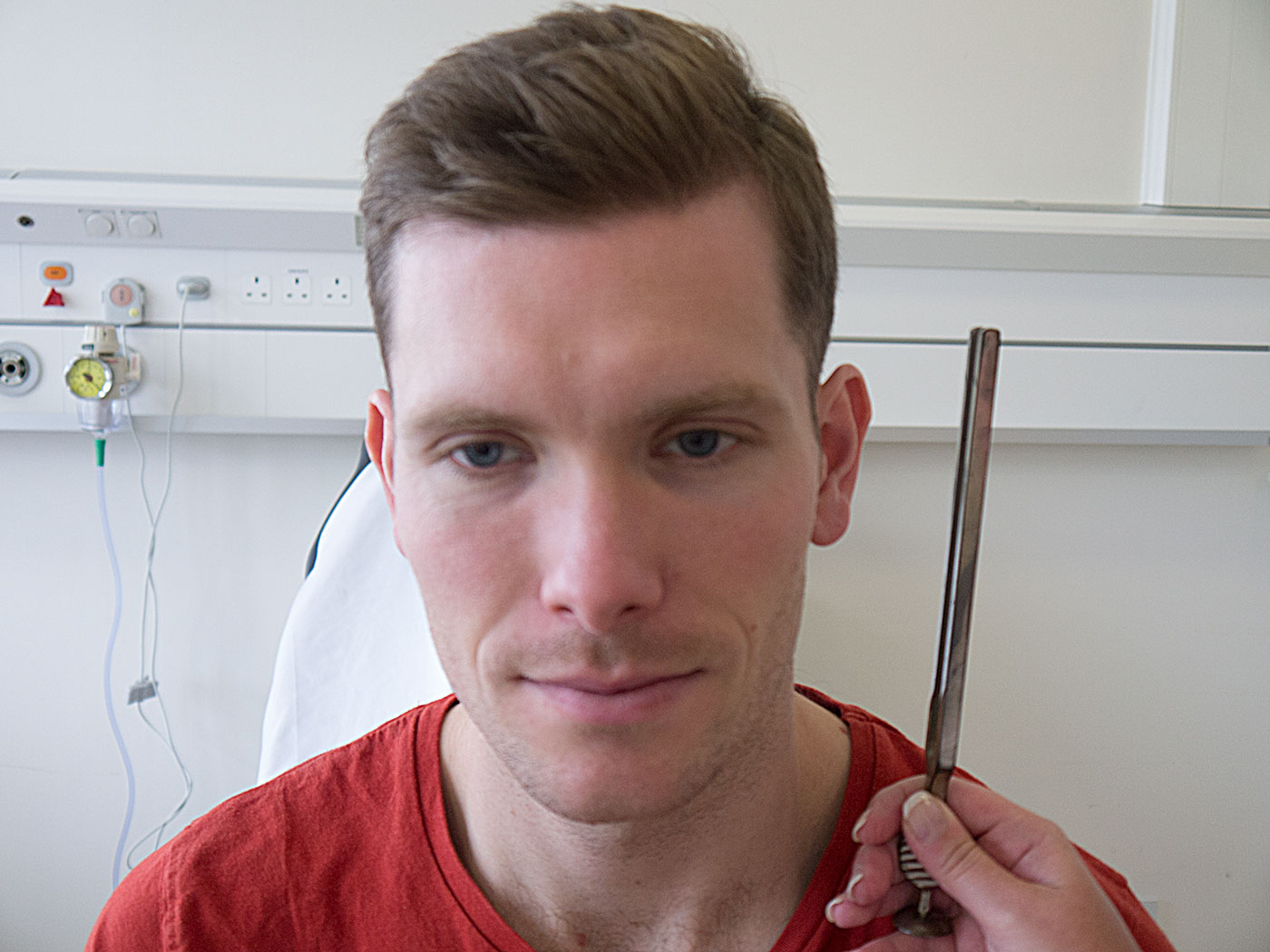
  • To carry out the Rinne test: Place a sounding tuning fork on the patient’s mastoid process and then next to their ear and ask which is louder. A normal patient will find the second position louder.
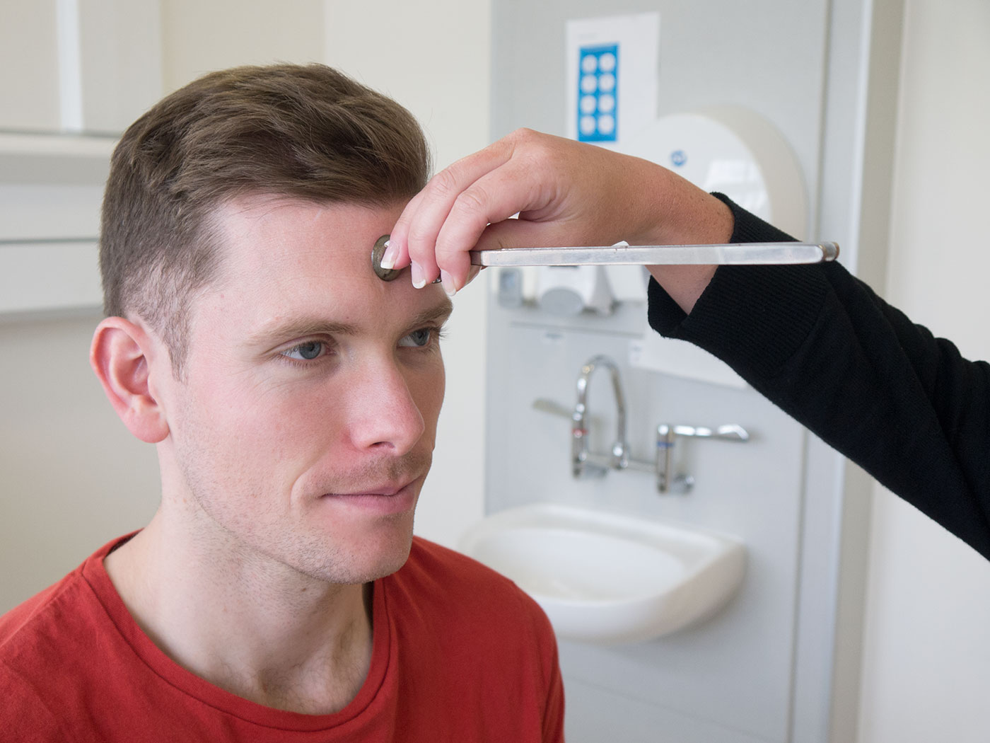
  • To carry out the Weber’s test: Place the tuning fork base down in the centre of the patient’s forehead and ask if it is louder in either ear. Normally it should be heard equally in both ears.Webers-test-place-the-tuning-fork-base-down-in-the-centre-of-the-forehead Rinne-test-place-tuning-fork-on-the-mastoid-process Rinne-test-place-tuning-fork-beside-the-ear
    Rinne's and Weber Test

CN 9 – Glossopharyngeal: 

  • Both motor and sensory and para sympathetic. 
  • Medulla oblongata, exits through the jugular foreman. 
  • Sensory: taste from posterior 1/3rd of the tongue, larynx, carotid sinus and body. 
  • Motor: pharyngeal muscle. 
  • Para symp: Salivary  glands. 
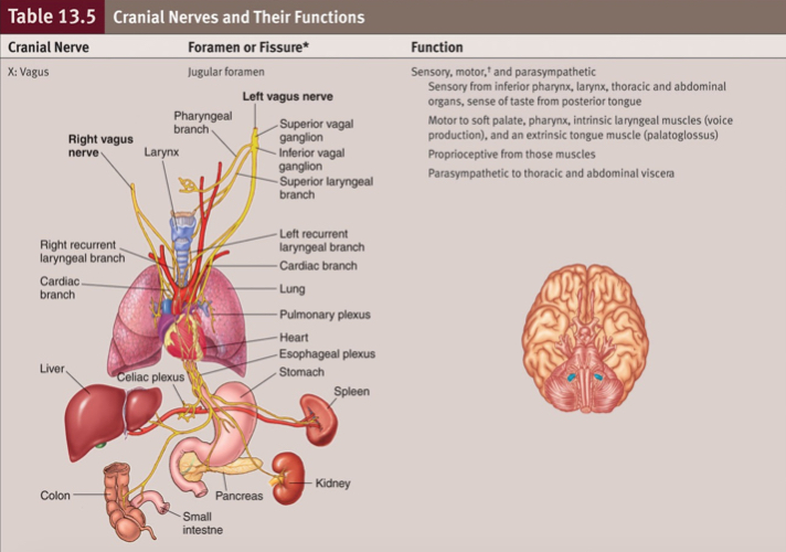
CN X – Vagus: 

  • Both motor and sensory. 
  • From the brain stem, through the jugular foreman to the viscera. 

Vagus nerve

  • Examination of CN IX & X: 
  • The Glossopharyngeal nerve: Provides sensory supply to the palate. It can be tested with the gag reflex or by touching the arches of the pharynx. 
  • The Vagus nerve (CN X):  provides motor supply to the pharynx. Asking the patient to speak gives a good indication to the efficacy of the muscles. The uvula should be observed before and during the patient saying “aah”. Check that it lies centrally and does not deviate on movement.
  • Dysfunction: Glossopharyngeal nerve & Vagus nerve: 
  • Damage results usually from surgery, cancer, aortic arch aneurysms. 
  • Dysfunction:
  • Dysphagia (difficulty swallowing) and dysarthria (difficulty talking) are caused by injury to the nuclei of the glossopharyngeal and vagus nerves.
  • Aphonia (inability to speak through disease of or damage to the larynx or mouth) or weak/hoarse voice are caused by injury to the vagus nerve. 

CN XI – The Accessory Nerve:

  • Motor. 
  • Medulla oblongata, jugular foramen and foramen magnum. 
  • SCM and Trapezius muscles, pharynx and soft palate. 
  • Spinal part prone to injury as it travels in the posterior lateral portion of the cervical spine. 
  • Examination: 
  • Cranial part – Testing the SCM. – resisted sidebending and rotation. 
  • Spinal part – Testing the trapezius. – resisted shoulder shrug. 

CN XII – Hypoglossal Nerve:

  • Motor.
  • Origin is the medulla oblongata, exiting out of the hypoglossal canal. 
  • Supplies all the intrinsic muscles and all extrinsic (except one) of the tongue, and the Styloglossus, Geniglossus, Hyoglossus.

Image472


  • Examination: 
  • Observe the tongue for any signs of wasting or fasciculations.
  • Ask the patient to stick their tongue out and move left, right, up, down. If the tongue deviates (on protrusion) or can’t complete to either side, it suggests a weakening of the muscles on that side. 

Thank you for reading! I hope you found this helpful! If you have anything to add, please let me know!

Like, Share, Comment, Follow.

Abdominal Examination

Abdominal Examination For OS4.


Exam Flow:

  • Inspection of the hands, face, tongue, chest and neck.
  • Radial pulse.
  • Palpation. – The 5 F’s.
    • + AAA.
    • + Superficial palpation.
  • Auscultation.
  • Palpation.
    • + Deeper palpation – organomegaly.
      • + Liver.
      • + Spleen.
      • + Kidneys.
      • + Bladder. (optional).
  • Percuss.

Inspection of the hands, face, eyes, tongue, neck and chest:

  • First of all, have the patient seated on the couch, laying down – exposed from the waist up. Throughout this exam, always keep in mind the 5 F’s:
  • Fat.
  • Fluid.
  • Fetus.
  • Flatus. (Gas).
  • Faeces.

Hands: 

  • Koil-onychia:
  • Also known as spoon nails, is a nail disease that can be a sign of hypochromic anaemia, especially iron-deficiency anaemia.
  • Leuk-onychia: 
  • Known as white nails or milk spots, is a medical term for white discoloration appearing on nails. Due to air bubbles present between the nail and its bed. Also Hypoalbumineamia.
  • Clubbing:
  • An abnormal widening and thickening of the finger tips and nails. 
  • + Crohn’s disease. (Type of inflammatory bowel disease (IBD) that may affect any part of the gastrointestinal tract from mouth to anus, commonly effecting the colon and ileum. Associated with ulcers and fistulae). 
  • + Ulcerative colitis. (A form of inflammatory disease (IBD) of the colon, the largest part of the large intestines. That includes characteristic ulcers, or open sores). 
  • + Coeliac disease. (Small intestine is hypersensitive to gluten, leading to difficulty in digesting food). 
  • To test for clubbing, use the Schamroth’s Window Test.
  • Asterixis (Flapping tremor): 
  • Quite slow frequency, whole hand tilts forwards – a result of encephalopathy (brain function disease) caused by urea.
  • Palmar erythema: 
  • A reddening of the thenar and hypothenar eminences.
  • “Corn Beef Hands”.
  • Chronic liver disease.
  • Pregnancy.
  • Nicotine staining. 
  • Dupuytren’s contracture: 
  • Alcoholic liver disease.
  • Known as Vikings Disease.
  • Is a connective tissue disorder – a fixed flexion contracture of the hand due to palmar fibromatosis (refers to a group of benign (non-harmful) soft tissue tumours).

Eyes: 

  • Check the conjunctiva (mucous membrane that covers the front of the eye and lines the inside of the eyelids) to check for Pallor    –> anaemia.
  • Check the Sclera (White outer layer of the eyeball) for jaundice (excess pigment bilirubin and typically caused by obstruction of the bile duct, liver disease, or excessive breakdown of RBC’s).
  • Xanthelasma. 

Mouth: 

  • Patient opens, look at the inside of the mouth, inside of cheeks for any ulcers –> Chron’s disease (Type of inflammatory bowel disease (IBD) that may affect any part of the gastrointestinal tract from mouth to anus, commonly effecting the colon and ileum. Associated with ulcers and fistulae). 
  • Look at the tongue.
    • + Fat and red = possible sign of anaemia. 
  • Side of the mouth where the lips meet.
    • + Angular Stomatitis, Inflammation corners of the mouth commonly unilaterally but can be bilateral. Commonly linked to candidiasis (oral thrush). 

450px-Angular_Cheilitis

Neck: 

  • Palpate the Virchow’s Node in the upper left supraclavicular fossa, this node drains the thoracic duct. Enlargement or Troisier’s sign may suggest metastatic cancer from either the abdomen or lungs.

Radial Pulse Check:

  • There is some argument as to whether this should be performed or not in an abdominal exam.
  • Good indicator of Sepsis (Harmful bacteria and their toxins in tissue) or Thyroid Disease.
  • Rate or bounding.
  • Rate is increased with asthma patients.
  • At the wrist, lateral to the flexor carpi radialis.
  • Three fingers over one artery, assess rate, rhythm and volume.
  • Count over 15 seconds and multiply by 4.
  • Then palpate both radial pulses at the same time to assess volume differences.
  • Palpate the radial and ipsilateral femoral pulse (radio-femoral delay).

Next step is to get the patient to lay down flat on the couch from the currently seated position.

Chest: 

  • Spider Naevi. A cluster of minute red blood vessels visible under the skin, occurring typically during pregnancy or as a symptom of certain diseases (e.g. Alcoholic cirrhosis, Liver cirrhosis but can be caused by any condition that results in increased levels of oestrogen). 5 or more = significant.

Spider Naevi

  • In males, check for Gynaecomastia: (Enlargement of a man’s breasts. Hormone imbalance).
  • Both of these are characteristics of Liver Pathology.

Now just have a general over look of the abdomen: 

  • Scars.
  • Masses.
  • Pulsations.
  • Abdominal distension – fluid/air that has built up to go beyond the normal girth of the stomach and waist. Underlying disease or dysfunction in the body rather than an illness in its own right. “Bloated Feeling”.

Auscultation:

  • Auscultation for bowel sounds may be carried out before percussion and palpation due to adverse effect that these procedures may have on the sound from the bowels.
  • Listen with the diaphragm around the quadrants for up to 30 seconds.
  • Listen for arterial bruits over the aorta. They may also arise from stenosis of renal arteries.
  • Listen for friction rubs over liver and spleen.
  • High pitched may indicate bowel obstruction. Sounds like water being poured from one cup to another. 
  • Absence of sounds may be also be caused by peritonitis (inflammation of the peritoneum). 

liver-location-in-abdomen-quadrants


Palpation:

  • Firstly palpate the abdominal aorta checking for AAA.
  • Palpation of the abdomen should be performed in a systematic way using the 9 named segments of the abdomen:
    • + Right and left hypochondrium,
    • + Right and left lumbar flanks,
    • + The umbilical area,
    • + The hypogastric,
    • + The right and left iliac regions.

    Abdomial Exam

  • Initial examination should be superficial using one hand. Place the hand flat over each area and flex at the metacarpophalangeal joints. You should feel whether the abdomen is soft but you should always be looking at the patient’s face for any signs of pain. – hedgehog hand.
  • Once all 9 areas have been examined superficially, you should move on to examine deeper.
  • A deeper exam is performed with two hands, one on top of the other again flexing at the MCP joints.
  • Use Mc’Burney’s Point, add pressure, if pain = possibility of appendicitis.

screen_shot_2013-09-11_at_24101_pm-1410ebc38680d3f2080

  • Murphy’s sign can be elicited by placing your examining fingers over the gallbladder area and then asking the patient to take a deep breath.
  • If Murphy’s sign is positive, there will be sudden reaction of the pain on inspiration and inspiration will be inhibited.

Surface_projections_of_the_organs_of_the_trunk


Organomegaly:

Liver, Spleen and Kidneys:

  • Palpation for the liver and spleen is similar, both starting in the right iliac fossa.
  • For the liver: press upwards towards the right hypochondrium. You should try to time the palpation with the patient’s breathing – in as this presses down on the liver If nothing is felt you should move towards the costal margin. Should feel soft but firm. (Fish tongue). 
  • Palpating for the spleen: Start well below the left costal margin. Ask patient to breath deeply, feel for splenic edge as it descends on inspiration. Not normally palpable. 
  • To feel for the kidneys: Right lower than Left. General population can’t be palpable. Place one hand under the patient L1/L2 in the lateral region and the other hand on top. You should then try to bring the kidney between the two hands by bring up the inferior hand and pushing the superior hand down. In the majority of people the kidneys are not palpable, but they maybe in thin patients who have no renal pathology.
  • Kidney palpation can also be done seated too. Palpate the renal angle for tenderness. Gently percuss with closed fist “Kidney Punch.” Abdominal Contents

Percussion:

  • Percussion over the abdomen is usually resonant (deep and clean sound), if there is a dull sound it usually means solid or liquid. 
  • Percussion allows you to determine if abdominal distension is because of solid or cystic tumours, ascites (the accumulation of fluid in the peritoneal cavity, causing abdominal swelling), or gas.
  • Fullness of the flanks may be the first indication of ascites. 
  • Percussion for shifting dullness: 
  • Supine. Percuss from the umbilical region moving down towards one side.
  • When the sound becomes dull, mark the spot (or keep your finger there) and ask the patient to move on to the opposite side. Give a short while for the fluid to sink and percuss again.
  • If the marked spot now becomes resonant that is a positive sign of ascites .
  • Percuss back down towards the umbilicus until dullness is reached again. Repeat on the other side.
  • Eliciting a fluid thrill: 
  • With one hand on the patient’s flank, flick the skin over the other flank using a finger. If an impulse or ‘fluid thrill’ is felt, this indicates a positive sign.
  • However, to be certain, you should repeat the examination with the patient’s hand along their midline in the sagittal plane to dampen any possible thrill transmitted by the abdominal wall.

It can be considered to do that auscultation part of this exam again, to check for possible changes in the gut due to all of the previous changes.

Thank you for reading! I hope you found this helpful! If you have anything to add, please let me know!

Like, Share, Comment, Follow.

Respiratory Examination

Respiratory Exam For OS4


Exam Flow:

  • Inspection of the hands, face and tongue.
  • Radial pulse (bounding pulse, an extremely strong and powerful pulse).
  • Inspect the Jugular Venous Pressure (JVP).
  • Front of the chest:
    • + Chest shape. –  Reduced chest expansion, LBP referring into glutes = possible ankylosing spondylitis. Also affects the costal cartilage.
    • + Breathing pattern. Around 14 per minute.
    • + Palpate the Trachea / Precordium (area of the chest that is immediately over the heart) / Apex.
    • + Palpate the chest expansion.
    • + Percuss / compare.
    • + Assess TVF / Compare. (Tactile Vocal Fremitus), a vibration transmitted through the body.
    • + Auscultate / Compare.
    • + Assess VR / Compare. (Vocal Fremitus).
  • Then perform the same things but on the back of the chest wall.

Inspection of the hands, nails, eyes, face and tongue.

General: 

  • Pallor, scars, fullness of the supraclavicular fossa, accessory breathing muscle patterns,
  • Chest deformities:
    • + Pectus Excavatum is the most common congenital deformity of the anterior wall of the chest, in which several ribs and the sternum grow abnormally. This produces a caved-in or sunken appearance of the chest. It can either be present at birth or not develop until puberty.
    • + Pectus Carinatum also called pigeon chest: is a deformity of the chest characterized by a protrusion of the sternum and ribs.

Hands:

  • Clubbing: 
  • Common cause:
    • + Bronchiectasis. – Abnormal widening of airways —> build up of excess mucous.
    • + Lung cancer. 
    • + Idiopathic pulmonary fibrosis. – Poorly understood causes. Scarring of lungs.
    • + Cyanotic congestive heart disease. 
    • + Infective endocarditis –  is a form of endocarditis, inflammation of the endocardium, the inner tissue of the heart (such as its valves) caused by infectious agents (pathogens).
  • To test for clubbing, use the Schamroth’s Window Test.
  • Peripheral cyanosis: 
  • Low O2 saturation.
    • + Right sided heart failure.
  • Peripheral Oedema: 
  • Swelling of tissues, usually in the lower limbs, due to the accumulation of fluids.
  • Commonly associated with ageing.
  • Other conditions, including:
    • + Congestive heart failure.
    • +Trauma.
    • + Alcoholism,
    • + Altitude sickness,
    • + Pregnancy.
    • + Hypertension.
    • + Right sided heart failure.
  • Palmar erythema: 
  • A reddening of the thenar and hypothenar eminences.
    • + Portal hypertension.
    • + Pregnancy.
    • + Chronic liver disease.
  • “Corn Beef Hands”.
  • Flapping Tremor / Asterixis:
    • + CO2 retention – COPD (chronic obstructive pulmonary disease, involving constriction of the airways and difficulty or discomfort in breathing).
    • + Liver failure. (Acute not so much – Paracetamol overdose).
    • + Can also be a feature of Wilson’s Disease: (copper accumulation disease in the tissues, marked by a copper ring around the iris, especially when neurological symptoms are present). 
  • +’ve = 20-30 second of hands in front of the patient, on a straight level, hands will begin to flap.
  • Face swelling: 
  • Think JVP = cause of face swelling.
  • Look for nicotine stains, drug use signs in this part of the exam too.

Face and Eyes: 

  • Ask the patient to poke out their tongue, to check for colour.
  • Cyanosis: 
  • Low O2 saturation.
  • Anemia: 
  • A condition in which there is a deficiency of red cells or of haemoglobin in the blood, resulting in pallor and weariness. Haemoglobin (made up of iron and protein) are responsible for transporting oxygen around our respiratory system, but if we do not have enough iron in our blood the haemoglobin become weak so are unable to transport oxygen around our body, which could leave us breathless, weak and less energetic.
  • Look for increased SCM tone.
  • Palpate the Virchow’s Node:
  • In the upper left supraclavicular fossa, this node drains the thoracic duct.
  • Enlargement or Troisier’s sign may suggest metastatic cancer from either the abdomen or lungs. 
  • Horner’s syndrome: 
  • A condition marked by a contracted pupil, drooping upper eyelid, and local inability to sweat on one side of the face, caused by damage to sympathetic nerves on that side of the neck. Possible Apical lung tumour.
    • + Same side face.
    • + Small pupil (miosis).
    • + Droop eyelid (partial ptosis). Superior tarsal muscle.
    • + Reduced sweat (anhidrosis).
  • Cause – problem of the 1st, 2nd, 3rd order neurones of the sympathetic nervous system.
  • Stroke / Lung / Thyroid Tumour / Trauma.
  • C8-T1 ANS block of sympathetic nerve to the eye.  hornerHorners Syndrome

Radial Pulse Check:

  • Rate or bounding.
  • Rate is increased with asthma patients.
  • At the wrist, lateral to the flexor carpi radialis.
  • Three fingers over one artery, assess rate, rhythm and volume.
  • Count over 15 seconds and multiply by 4.
  • Then palpate both radial pulses at the same time to assess volume differences.
  • Palpate the radial and ipsilateral femoral pulse (radio-femoral delay).
  • Tachycardia suggests significant respiratory difficulty. Lung cancer can cause atrial fibrillation. A large pneumothorax or a tension pneumothorax can cause pulsus paradoxus (is an abnormally large decrease in systolic blood pressure and pulse wave amplitude during inspiration).

JVP Check:

  • Recline the patient to 45 degrees, ensure the neck is relaxed and head facing away, look between the two sternocleidomastoid heads for a pulsation. If there is a pulsation, you need to determine whether it actually is the JVP. The JVP is non-palpable, obliterated by compressing, has double waveform and is exaggerated with hepatojugular reflex.
  • Once the JVP is confirmed, note its height from the sternal angle (shouldn’t be more than 3-4cm).

Jugular-Venous-Pressure-web-large(800x600)

Causes of a raises JVP: 

  • Heart failure.
  • Constrictive pericarditis (JVP increases on inspiration – called Kussmaul’s sign).
  • Cardiac tamponade.
  • Fluid overload, eg renal disease.
  • Superior vena cava obstruction (no pulsation).

Palpate:

Chest Expansion

chest_exp1341276832524

  • Chest expansion:
  • Feeling for fluidity of movement and if opening properly.
  • Usual chest expansion in an adult is 4-5 cm and should be symmetrical.
  • Symmetrical reduction:
  • Overinflated lungs:
    • + Bronchial asthma – chronic inflammatory disease of the airways characterised by variable and recurring symptoms, reversible airflow obstruction and bronchospasm. Common symptoms include wheezing, coughing, chest tightness, and shortness of breath (SOB).
    • + Emphysema – a condition in which the air sacs of the lungs are damaged and enlarged, causing breathlessnessSmokers. Air is abnormally present within the body tissues.
  • Stiff lungs:
    • + Pulmonary fibrosis – Scarring in the lung tissue. Leading to serious breathing problems. Scar formation, the accumulation of excess fibrous connective tissue (the process called fibrosis), leads to thickening of the walls, and causes reduced oxygen supply in the blood. Patients suffer from SOB.
    • + Ankylosing spondylitis – Type of spinal arthritis. Bamboo spine.
  • Asymmetrical reduction of chest wall expansion:
  • Absent expansion:
    • + Empyema – Pus in a cavity, especially in the pleural cavity. 
    • + Pleural effusion – Excess fluid that accumulates in the pleural cavity. Various kinds = hemothorax (blood), urinothorax (urine), pyothorax (pus). Pneumothorax is the accumulation of air in the pleural space.
  • Reduced expansion:
    • + Pulmonary consolidation – Lung tissue that has filled with liquid, a condition marked by induration (swelling or hardening).
    • + Pneumothorax –  Accumulation of air in the pleural space. Collapsed lung.

Diagram_showing_a_build_up_of_fluid_in_the_lining_of_the_lungs_(pleural_effusion)_CRUK_054

  • Trachea position: 
  • The trachea may be pulled to one side by unilateral upper lobe fibrosis or collapse. Upper lobe fibrosis usually suggests tuberculosis in the past. Lung cancer is causing blockage of a main bronchus is usually responsible for upper lobe collapse. The trachea may also be displaced by a tension pneumothorax or by large pleural effusion, which would push the trachea to the opposite side. 

Trachea Position


 Percussing:

Purcuss

  • This is a very time-consuming skill, taking a lot of practice that will develop over time. The trick is to keep your palpation of the intercostal spaces light, and your percussing hand loose, but fingers firm.
  • Remember to get the patient to hug themselves to protract the scapulas out of the way – posterior percussing.
  • A hyper-resonant sound suggests hyperinflation or a pneumothorax.
  • A dull sound is easier to distinguish from normal. It may suggest collapse or consolidation, or a pleural effusion.

Percuss - Posterior Percuss Anterior

Vocal Fremitus:

  • This is where you use the ulnar borders of the hands on the areas in the picture, as the patient begins to say the number 99 as and when instructed. You are meant to be feeling for the vibrations that are caused as they move through the thorax. Tactile vocal fremitus is increased over areas of consolidation and decreased or absent over areas of effusion or collapse.

Vocal Resonance:

  • This is done the exact same way as the Fremitus, but using the stethoscope to listen for the vibrations.
  • This technique allows discrimination between dullness to percussion from pleural effusion and that from consolidation. It again reflects the properties of normal and abnormal lung for transmitting and filtering sound.
  • Auscultate over the chest in a systematic fashion while the patient says “ninety nine” repeatedly.
  • As previously mentioned, normal lung transmits lower and filters higher frequency sound leading to the numbers being indistinct and reverberating in quality.
  • Consolidated lung will transmit the higher frequencies and the numbers will sound far more distinct and with a vague buzzing quality, like speech after the inhalation of helium.
  • In the case of pleural disease such as effusion the vocal resonance will simply be much reduced in intensity or absent.

 Auscultate:

  • Same sites as the percussing technique.

What to listen for? – Hyperlinks.


  • Crackles (Rough) – Use Diaphragm:
  • Heard during inspiration. Louder, lower pitched and last longer than Fine.
  • Excessive fluid in the lungs.

  • Wheeze – Use Diaphragm: 
  • Can be high or low pitched, the proportion of the respiratory cycle occupied by the wheeze correlates to the degree of airway obstruction.
  • Narrowing of the airways.

  • Rhonchi – Use Diaphragm: 
  • Low pitched wheeze in both inspiration and expiration. Rhonchi occurs in the bronchi and heard over the chest wall not alveoli.
  • Usually cleared with coughing.

  • Pleural Rubs – Use Diaphragm:
  • Continuous or Discontinuous, sounds like creaking or grating sound or like walking on fresh snow. Coughing will not clear.
  • Inflamed or roughened pleural surfaces rubbing against one another.

  • Heart auscultation: 

  • Mainly to detect heart abnormalities but severe lung disease may cause pulmonary hypertension and a loud P2.
  • Place the stethoscope over each of the 5 lobes of the lungs in turn, on the front and back of the chest. Ask the patient to take deep breaths in and out with their mouth open.
  • Normal breath sounds are called vesicular. They are described as quiet and gentle. There is usually no gap between the inspiratory and expiratory phase sounds.

  • Rhonchi (wheezes):

  • Musical sound heard on expiration. In severe cases they may be both inspiratory and expiratory. Imply narrowing of the airways.
  • The loudness of wheezing gives no indication of the severity of the condition.

  • Rales (sometimes called crackles):

  • Probably represent opening of small airways and alveoli.
  • They may be normal at the lung bases if they clear on coughing or after taking a few deep breaths.
  • Basal crackles are a classical feature of pulmonary congestion with left ventricular failure. They may be more diffuse in pulmonary fibrosis.

  • Bronchial breathing:

  • The sounds of bronchial breathing are generated by turbulent air flow in large airways (similar sounds can be heard in healthy patients by listening over the trachea.
  • Sounds are harsh and poor in nature. Unlike normal vesicular breath sounds, there is a gap between the inspiratory and expiratory phase sounds.
  • Bronchial breathing suggests consolidation or fibrosis, which permits the sound to be conducted more effectively to the chest wall.

  • Pleural rub:

  • A creaking sound caused by stiff pleural membranes such as with pleurisy.

  • Stridor:

  • Harsh inspiratory sound caused by partial obstruction of a large airway.

Thank you for reading! I hope you found this helpful! If you have anything to add, please let me know!

Like, Share, Comment, Follow.